Cosmetic
Targeted correction of the under-eye hollow with filler, fat grafting, or lower blepharoplasty options for the tear trough deformity.
Medically reviewed by Noel D. Saks, MDOculoplastic SurgeonLast updated June 2026
The tear trough is the depression that runs obliquely from the inner corner of the eye downward and outward along the junction between the lower eyelid and the cheek. Anatomically, it corresponds to the tear trough ligament—a true osteocutaneous attachment that tethers the skin to the maxillary bone along the medial orbital rim. Lateral to this, the depression continues as the palpebromalar groove, defined by the orbicularis retaining ligament. Together these structures create the shadowed crescent that patients describe as looking “tired,” “sunken,” or “hollow,” even when they feel well rested.
A prominent tear trough is one of the most common cosmetic concerns brought to an oculoplastic surgeon. Unlike fine lines or pigmentation, the tear trough is a structural problem—a topographic depression caused by a combination of ligamentous tethering, soft-tissue volume loss, and herniation of orbital fat above the groove. Because the anatomy is layered and unforgiving, treatment requires nuanced judgment about which combination of volume restoration, fat repositioning, or skin tightening will produce a natural, durable result.
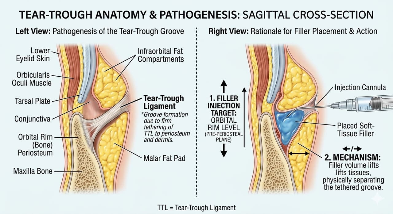
The tear trough deformity is rarely caused by a single factor. In most patients, three age-related changes converge:
Skin quality, pigmentation (melanin deposition or vascular show through thin skin), and bony anatomy (a negative vector with a recessed maxilla) all modify how the hollow appears. A careful exam differentiates true volume deficit from pseudohollowing caused by fat prolapse—a distinction that determines whether the right treatment is to add, reposition, or remove tissue.
Important: Filler placed into a hollow caused primarily by fat prolapse will worsen the bulge above and create a puffy, overcorrected appearance. Diagnosis matters more than product choice.
Hyaluronic acid (HA) filler is the least invasive option and the appropriate first-line treatment for many patients—particularly younger individuals with mild to moderate hollowing, minimal fat herniation, and good skin elasticity. The goal is to soften the transition between the lid and cheek, not to eliminate every shadow.
Filler is placed deep, on or just above periosteum, beneath the orbicularis oculi muscle. Superficial placement risks the Tyndall effect (a bluish discoloration from light scattering through HA), prolonged edema, and visible product contour. Most experienced injectors use a blunt cannula introduced from a lateral entry point, depositing small aliquots (typically 0.05–0.1 mL per pass) along the medial orbital rim. Total volume is usually 0.5–1.0 mL per side.
Lower-G′, less hydrophilic HAs (such as Restylane-L, Restylane Eyelight, or Belotero Balance) are preferred. Highly cross-linked, hydrophilic products designed for the cheek or jawline draw water aggressively and produce persistent under-eye puffiness that can last for years.
For a broader discussion of injectable products and placement principles, see our Fillers page.
Results are visible immediately, though mild swelling and occasional bruising take 1–2 weeks to settle. HA filler in the tear trough is notoriously long-lasting—often persisting 2–5 years or more because the area has minimal muscular movement and slow lymphatic clearance. The major advantage of HA is reversibility: hyaluronidase can dissolve unwanted product, often quickly, though more than one session is sometimes needed.
Autologous fat grafting (also called fat transfer or lipofilling) uses the patient’s own fat—harvested by gentle liposuction from the abdomen, thigh, or flank—to restore volume in the tear trough, lid–cheek junction, and midface. It is an excellent option for patients who want a more durable, biologically integrated correction and who have generalized periorbital and midface volume loss rather than an isolated trough.
Fat is harvested with a low-suction cannula, processed (decanted, centrifuged, or filtered) to concentrate viable adipocytes, and reinjected in microaliquots using small-gauge cannulas. Multiple tissue planes are layered to maximize graft survival and produce smooth contour. For the tear trough, fat is deposited deep, often combined with nanofat or microfat in more superficial planes for skin quality improvement.
Fat grafting is often combined with a midface lift or lower blepharoplasty when significant descent and fat prolapse coexist.
When the tear trough is created or worsened by prolapsing orbital fat, the most definitive correction is transconjunctival lower blepharoplasty with fat repositioning. Rather than excising the herniated fat (which can leave a hollow, skeletonized lower lid), the surgeon releases the orbicularis retaining ligament and tear trough ligament, then redrapes the patient’s own orbital fat across the orbital rim to fill the groove from within.
Through a hidden incision on the inner surface of the lower eyelid (no external scar), the medial, central, and sometimes lateral fat pads are mobilized as pedicled flaps. The arcus marginalis and tear trough ligament are released along the inferior orbital rim. The fat is then transposed over the rim into a subperiosteal or supraperiosteal pocket and secured with absorbable sutures. The result is a smooth, continuous lid–cheek transition—the bulge above and the hollow below are simultaneously corrected.
If the patient also has skin redundancy, fine wrinkling, or orbicularis hypertrophy, a small skin-pinch excision or a skin–muscle flap approach can be added. Adjunctive skin resurfacing with CO₂ or erbium laser further tightens crepey skin. See our pages on Blepharoplasty and Lasers for related options.
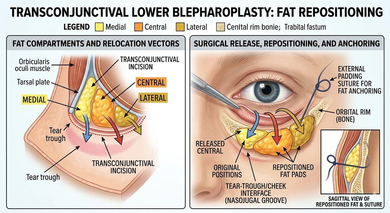
A generation ago, lower blepharoplasty meant removing fat. Many of those patients now present in their 60s and 70s with hollow, aged-looking lower lids that are difficult to revise. Modern oculoplastic technique preserves and repositions fat whenever possible—a philosophy strongly supported by ASOPRS fellowship training.
Choosing among filler, fat grafting, and surgery depends on patient age, anatomy, skin quality, goals, and tolerance for downtime. The table below summarizes typical matches.
| Patient Profile | Best First-Line Option | Why |
|---|---|---|
| 20s–30s, congenital trough, no fat bulge, good skin | HA filler | Minimally invasive, reversible, addresses pure volume deficit |
| 40s–50s, mild fat prolapse + hollowing, fair skin | Filler trial, then consider blepharoplasty | Reversible test of aesthetic goal before committing to surgery |
| 50s+, prominent fat bags, deep trough, lid–cheek step-off | Transconjunctival blepharoplasty with fat repositioning | Addresses cause (fat herniation) and fills trough simultaneously |
| Generalized midface deflation, thin skin, negative vector | Fat grafting ± midface lift | Restores volume across multiple zones, durable, autologous |
| Prior filler overcorrection, persistent puffiness | Hyaluronidase dissolution, then reassess | Old filler must be cleared before accurate diagnosis |
| Dark circles from pigmentation/vascularity, no hollow | Topicals, laser, PRP — not filler | Volume correction will not address chromatic causes |
Every treatment carries trade-offs. Discussing them honestly is part of informed consent.
Filler patients return to work the same day, with bruising masked by makeup after 48–72 hours. Fat grafting and lower blepharoplasty typically involve 7–10 days of visible swelling and bruising, with residual mild edema resolving over 6–12 weeks. Cold compresses, head elevation, and avoidance of strenuous activity for two weeks are standard. Final aesthetic results from surgery are best judged at 3–6 months.
Patients with significant Dry Eye Disease or Eyelid Laxity require additional planning before lower-lid surgery to prevent post-operative complications.
The lower eyelid is the most unforgiving region in facial aesthetics. Tissue is thin, the orbital septum is shallow, and small errors—a millimeter of malposition, a fraction of a milliliter of misplaced filler—produce visible, sometimes irreversible problems. For this reason, tear trough treatment is best entrusted to an oculoplastic surgeon with ASOPRS fellowship training. These surgeons spend two additional years after ophthalmology residency mastering eyelid anatomy, orbital surgery, and periocular aesthetics—and they can manage complications (vascular events, lid malposition, dry eye) that other injectors and surgeons may not be equipped to address.
When consulting, ask whether Dr. Saks performs the full spectrum of tear-trough treatments—filler, fat grafting, and surgical fat repositioning. A surgeon who only offers one option may steer you toward that option regardless of whether it is the best fit. The right plan starts with an honest assessment of your anatomy and goals.
Ready to explore your options?
Schedule a consultation with Noel D. Saks, MD to learn if this procedure is right for you.